What is 3D Mammography?
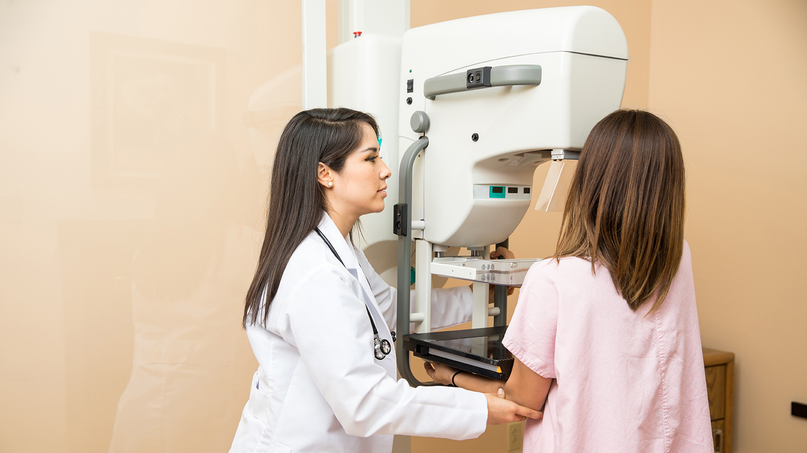
A tomosynthesis exam is very similar to a traditional mammogram and takes approximately the same amount of time. Just as with a digital mammogram, the technologist positions the patient then compresses the patient’s breast under a paddle and takes images from different angles.
Breast tomosynthesis uses high-powered computing to convert digital breast images into a stack of very thin layers or “slices”—building what is essentially a “3-dimensional mammogram.”
Increased Detection
Now, however, the radiologist can see breast tissue detail in a way never before possible. Instead of viewing all the complexities of your breast tissue in a flat image, the doctor can examine the tissue a millimeter at a time. Fine details are more clearly visible, no longer hidden by the tissue above and below.
During the tomosynthesis part of the exam, the X-ray arm sweeps in a slight arc over the breast, taking multiple breast images in just seconds. A computer then produces a 3D image of your breast tissue in one-millimeter layers.
Recognized as Providing Excellent Breast Healthcare

Having this advanced technology and the trained staff to support it has also qualified South County Health as a Pink Ribbon Facility—a digital imaging center recognized as providing excellent breast healthcare paired with exceptional commitment and support to the women of their community.
Locations
“My ultrasound technologist had a very gentle, compassionate demeanor that provided me with a sense of comfort. Her professional, warm personality gave me the assurance I was in a good place. ”
- Anonymous Patient