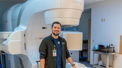
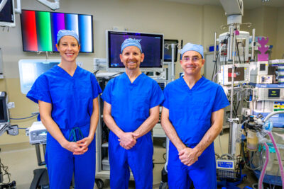
Find a Physician Browse
Find a Physician

Technology transforms diagnostics

Published 09/09/2019

Xray1Named for the man who invented it, the Roentgen X-ray was state-of-the-art technology in the 1920s. This simple imaging technique, discovered by German physicist Wilhelm Roentgen in 1895, had seen wide use in hospitals.

When the brick South County Hospital was built in 1925, it would include an X-ray machine.

According to the hospital's 1928 annual report, 114 patients were tested with X-rays totaling 231 exposed films. By 1939, that number had increased to 609 cases. They included “fluoroscopy,” a technology invented by Thomas Edison that employs X-rays to obtain moving pictures, such as of a beating heart.

In an effort to improve patient care with the new technology, the hospital hired a consulting radiologist, Dr. Russell R. Hunt, in November 1938. It also refurbished the X-ray room, and purchased “a new portable shock proof x-ray machine for bedside work,” The Narragansett Times reported. The cost: $550, of which Dr. John Paul Jones found a donor to cover all but $50.

Although imaging increased in sophistication, for the next 50 years the hospital would employ machines that changed little in principle since the new hospital had opened in 1925.

Addressing a community health concern

One of the earliest uses of the X-ray was to diagnose tuberculosis. Although the hospital had its own equipment, in 1945 a doctor and X-ray technologist from the Wallum Lake Sanitorium came to the hospital to hold a “chest clinic.” It is possible the war had disrupted the hospital's X-ray staffing.
The first major expansion of X-ray capacity came in August 1952, when the hospital's first addition, the Hazard Wing, opened. Besides 14 beds and expanded laundry facilities, the addition included a larger X-ray space.

By the time Donald L. Ford was hired to be the hospital's administrator, in 1958, South County still had only a part-time radiologist who came once a week. Dr. John J. Walsh recalled that in the early years doctors simply read their own X-rays rather than waiting for the radiologist to show up.

That year, the X-ray facilities themselves were judged “adequate and well equipped” by the consulting architects hired in preparation for the 1962 addition. After that addition was built, X-ray use increased along with the area's population, and the number of doctors on staff. In 1962, 5,839 X-rays were taken annually.

Like a camera

In 1971, Joyce MacManus joined the staff at South County Hospital, entering this “low-tech” world as an X-ray technician. The technicians were responsible for taking the X-ray and developing the film.

“Back then, you could compare it to the old manual cameras,” she said. “You had to set the F stop and the speed and all that …now it's all automatic.”

The ER also had a portable X-ray, but it was tethered by a long extension cord to a 220-volt outlet. Now, the device uses wireless technology, and the image comes up on a screen as the picture is taken.

The first improvement to imaging came with the introduction of ultrasound to the hospital in 1972. The images were grainy and in black and white, but provided real-time observation of beating hearts, helpful in detecting conditions such as valve abnormalities.

The “wave of the future”

The use of radioactive dyes to enhance imaging also began in the 1970s, leading to the formation of the hospital's next addition in 1978 – space for Nuclear Medicine and ultrasound.

But it would be 1985 before the hospital finally acquired a CT scan, and it would take a winding road to get there.

President Donald L. Ford was initially opposed to acquiring a CT scan, recalled Rudi Hempe, a Trustee who served on the Board from 1975 to 1992. Ford thought the technology was too expensive for the small community hospital, and that it would never receive a certificate of need from the state Department of Health. Instead, patients were referred to the larger regional hospitals or wait for the portable CT scan to show up once a month.

“You had these roving units in a trailer,” recalled Hempe, who served on the Trustees' equipment subcommittee. Eventually, he recalled, the committee realized that CT scans were the wave of the future, and Hempe was deputized to convince Ford.

“I said, 'Don, we're going to have to have our own CT scan,'” Hempe said. “ … and he said 'Never, never going to happen.' So we made a bet. A bottle of scotch.” He laughed. “I still have the bottle of scotch.”

In announcing its application for a certificate of need, the hospital wrote: “The CT Scanner is no longer considered a luxury that should be available only at large institutions.” The technology “provides detailed cross-sectional pictures of organs and bones never before achieved through conventional X-rays.”

The hospital noted that the CT scan eliminates the need for exploratory surgery and long hospitalizations, “and has proven itself to be effective in diagnosing cancer, stroke, trauma-related injuries, infection, as well as neurological and orthopedic disorders.”

The state granted the certificate of need, and the CT scan arrived in 1985. The price tag: $775,000. A small addition was built to house it.

“The number of scans performed daily has exceeded our expectations,” reported board President Albert C. Henry Jr. in the 1985 annual report.

That same year the hospital added a laser for microsurgery of the eye; a defibrillator and heart monitor; and a new cardiopulmonary unit, giving stress tests and electro-encephalograms.

South County Hospital also became the first in the state to own a two-dimensional color Doppler ultrasound. A new age of technology was on the horizon.

Xray2

Next: Advances in cardiology and surgery will transform patient care.